Will miscarriage care remain available?

When you first learned the facts about pregnancy — from a parent, perhaps, or a friend — you probably didn’t learn that up to one in three ends in a miscarriage.
What causes miscarriage? How is it treated? And why is appropriate health care for miscarriage under scrutiny — and in some parts of the US, getting harder to find?
What is miscarriage?
Many people who come to us for care are excited and hopeful about building their families. It’s devastating when a hoped-for pregnancy ends early.
Miscarriage is a catch-all term for a pregnancy loss before 20 weeks, counting from the first day of the last menstrual period. Miscarriage happens in as many as one in three pregnancies, although the risk gradually decreases as pregnancy progresses. By 20 weeks, it occurs in fewer than one in 100 pregnancies.
What causes miscarriage?
Usually, there is no obvious or single cause for miscarriage. Some factors raise risk, such as:
- Pregnancy at older ages. Chromosome abnormalities are a common cause of pregnancy loss. As people age, this risk rises.
- Autoimmune disorders. While many pregnant people with autoimmune disorders like lupus or Sjogren’s syndrome have successful pregnancies, their risk for pregnancy loss is higher.
- Certain illnesses. Diabetes or thyroid disease, if poorly controlled, can raise risk.
- Certain conditions in the uterus. Uterine fibroids, polyps, or malformations may contribute to miscarriage.
- Previous miscarriages. Having a miscarriage slightly increases risk for miscarriage in the next pregnancy. For instance, if a pregnant person’s risk of miscarriage is one in 10, it may increase to 1.5 in 10 after their first miscarriage, and four in 10 after having three miscarriages.
- Certain medicines. A developing pregnancy may be harmed by certain medicines. It’s safest to plan pregnancy and receive pre-pregnancy counseling if you have a chronic illness or condition.
How is miscarriage diagnosed?
Before ultrasounds in early pregnancy became widely available, many miscarriages were diagnosed based on symptoms like bleeding and cramping. Now, people may be diagnosed with a miscarriage or early pregnancy loss on a routine ultrasound before they notice any symptoms.
How is miscarriage treated?
Being able to choose the next step in treatment may help emotionally. When there are no complications and the miscarriage occurs during the first trimester (up to 13 weeks of pregnancy), the options are:
Take no action. Passing blood and pregnancy tissue often occurs at home naturally, without need for medications or a procedure. Within a week, 25% to 50% will pass pregnancy tissue; more than 80% of those who experience bleeding as a sign of miscarriage will pass the pregnancy tissue within two weeks.
What to know: This can be a safe option for some people, but not all. For example, heavy bleeding would not be safe for a person who has anemia (lower than normal red blood cell counts).
Take medication. The most effective option uses two medicines: mifepristone is taken first, followed by misoprostol. Using only misoprostol is a less effective option. The two-step combination is 90% successful in helping the body pass pregnancy tissue; taking misoprostol alone is 70% to 80% successful in doing so.
What to know: Bleeding and cramping typically start a few hours after taking misoprostol. If bleeding does not start, or there is pregnancy tissue still left in the uterus, a surgical procedure may be necessary: this happens in about one in 10 people using both medicines and one in four people who use only misoprostol.
Use a procedure. During dilation and curettage (D&C), the cervix is dilated (widened) so that instruments can be inserted into the uterus to remove the pregnancy tissue. This procedure is nearly 99% successful.
What to know: If someone is having life-threatening bleeding or has signs of infection, this is the safest option. This procedure is typically done in an operating room or surgery center. In some instances, it is offered in a doctor’s office.
If you have a miscarriage during the second trimester of pregnancy (after 13 weeks), discuss the safest and best plan with your doctor. Generally, second trimester miscarriages will require a procedure and cannot be managed at home.
Red flags: When to ask for help during a miscarriage
During the first 13 weeks of pregnancy: Contact your health care provider or go to the emergency department immediately if you experience
- heavy bleeding combined with dizziness, lightheadedness, or feeling faint
- fever above 100.4° F
- severe abdominal pain not relieved by over-the-counter pain medicine, such as acetaminophen (Tylenol) or ibuprofen (Motrin, Advil). Please note: ibuprofen is not recommended during pregnancy, but is safe to take if a miscarriage has been diagnosed.
After 13 weeks of pregnancy: Contact your health care provider or go to the emergency department immediately if you experience
- any symptoms listed above
- leakage of fluid (possibly your water may have broken)
- severe abdominal or back pain (similar to contractions).
How is care for miscarriages changing?
Unfortunately, political interference has had significant impact on safe, effective miscarriage care:
- Some states have banned a procedure used to treat second trimester miscarriage. Called dilation and evacuation (D&E), this removes pregnancy tissue through the cervix without making any incisions. A D&E can be lifesaving in instances when heavy bleeding or infection is complicating a miscarriage.
- Federal and state lawsuits, or laws banning or seeking to ban mifepristone for abortion care, directly limit access to a safe, effective drug approved for miscarriage care. This could affect miscarriage care nationwide.
- Many laws and lawsuits that interfere with miscarriage care offer an exception to save the life of a pregnant patient. However, miscarriage complications may develop unexpectedly and worsen quickly, making it hard to ensure that people will receive prompt care in life-threatening situations.
- States that ban or restrict abortion are less likely to have doctors trained to perform a full range of miscarriage care procedures. What’s more, clinicians in training, such as resident physicians and medical students, may never learn how to perform a potentially lifesaving procedure.
Ultimately, legislation or court rulings that ban or restrict abortion care will decrease the ability of doctors and nurses to provide the highest quality miscarriage care. We can help by asking our lawmakers not to pass laws that prevent people from being able to get reproductive health care, such as restricting medications and procedures for abortion and miscarriage care.
About the Authors

Sara Neill, MD, MPH, Contributor
Dr. Sara Neill is a physician-researcher in the department of obstetrics & gynecology at Beth Israel Deaconess Medical Center and Harvard Medical School. She completed a fellowship in complex family planning at Brigham and Women's Hospital, and … See Full Bio View all posts by Sara Neill, MD, MPH 
Scott Shainker, DO, MS, Contributor
Scott Shainker, D.O, M.S., is a maternal-fetal medicine specialist in the Department of Obstetrics and Gynecology at Beth Israel Deaconess Medical Center (BIDMC). He is also a member of the faculty in the Department of Obstetrics, … See Full Bio View all posts by Scott Shainker, DO, MS

Ready to give up the lead vest?

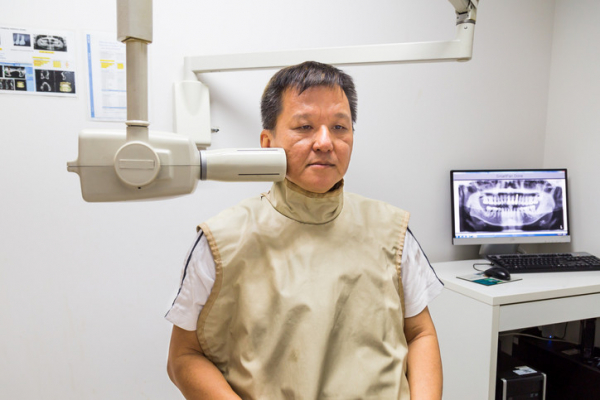
At a dental appointment last month, I spotted a lead vest hanging unassumingly on the wall of the exam room as soon as I walked in. “Still there, but now obsolete,” I thought.
I’d just learned about new guidelines from the American Dental Association (ADA) saying lead vests and thyroid collars that cover the neck are no longer needed during dental x-rays. But they’d been a fixture of my dental experiences — including many cavities, four root canals, a tooth extraction, and two crowns — for my entire life. What changed, and could I feel safe without the vest?
Why were lead vests used in past years?
Lead vests and thyroid collars have been worn by countless Americans during dental x-rays over the years. They’ve been in use for far longer than my lifetime — about 100 years. The heavy apron-like shields are placed over sensitive areas, including the chest and neck, before the x-rays are taken.
“I haven’t worn a lead apron in the last 10 or 15 years — unless a dentist insists I put it on — because I know it isn’t needed,” says Dr. Bernard Friedland, an associate professor of oral medicine, infection, and immunity at Harvard School of Dental Medicine.
What has changed about dental x-rays?
When lead vests and thyroid collars were first recommended, x-ray technology was much less precise. But the technology has evolved significantly over the last few decades in ways that dramatically improve patient safety:
- Digital x-rays enable far smaller radiation doses, reducing radiation exposure and the risks associated with higher doses, such as cancer. “The doses used in dental radiology are negligibly small now. If you go to the dentist today for a full series of mouth x-rays that are taken with a digital sensor, the total exposure time is just over five seconds,” explains Dr. Friedland, an expert in oral radiology. “A hundred or so years ago, that exposure time would have been many minutes.”
- The small size of today’s x-ray beam significantly reduces radiation “scatter” and restricts the beam size to only the area needing to be imaged. This protects patients from radiation exposure to other parts of the body.
A less-recognized strike against using lead vests and thyroid collars is their ability to get in the way. They may block the primary x-ray beam, preventing dentists from capturing needed images. This quirk can lead to repeat imaging and unnecessary exposure to additional radiation. This is more likely to occur with panoramic x-rays.
The gear may also spread germs, Dr. Friedland notes. Although disinfected, it’s not sterilized between uses. “There’s a risk of spreading bacteria and viruses,” he says. “To me, that’s also an issue and another reason I don’t want to use one on myself.”
Who no longer needs the shields?
No one does — even children, who presumably have a long life of dental x-rays in front of them. The new recommendations apply to all patients regardless of age, health status, or pregnancy, the ADA says.
The recommendation to discontinue lead vests has been a long time in the making. In fact, the ADA isn’t the first professional organization to propose it. The American Association of Physicists in Medicine did so in 2019, followed by the American College of Radiology in 2021 and the American Academy of Oral and Maxillofacial Radiology in 2023.
Are some people confused or concerned about the no-lead-vest policy?
Yes. The new guidelines are bound to draw confusion and fear, Dr. Friedland says. Some people may even insist on continuing to wear a lead vest during x-rays.
“A big problem is that people’s perception of risk is very skewed,” he says. “Some people, you’ll never convince.”
People are likely to feel more comfortable if the practice is uniformly adopted by dentists. However, the ability to implement this change may hinge partly on public response. And it could take a while to fully adopt.
“I think the public is going to have more say on this than dentists,” Dr. Friedland says. “It might take a generation to make this change, maybe longer.”
Still concerned about the new recommendations?
If you have lingering concerns about the new recommendations, talk to your dentist.
And ask if dental x-rays are necessary to proceed with your diagnosis or treatment plan. Sometimes it’s possible to take fewer x-rays — such as bitewing x-rays of the upper and lower back teeth only — or to use certain types of imaging less frequently. Even with far safer x-ray conditions, dentists should be able to justify that the information from images is integral to diagnose problems or improve care, Dr. Friedland says.
It’s worth noting that the dose of radiation, while far lower than in the past, varies with the type of imaging and which parts of the jaw are being imaged. For example, the digital dental x-rays mentioned above involve less radiation than conventional dental x-rays. Either panoramic dental x-rays, or 3-D dental x-rays taken with a CBCT system that rotates around the head, typically involve more radiation than conventional dental x-rays.
Whenever possible, dentists should use images taken during previous dental exams, according to the ADA. “If I don’t need an x-ray, I don’t get one,” says Dr. Friedland. “I’m not cavalier about it. I also use technical parameters that keep the x-ray dose as low as reasonably possible.”
About the Author

Maureen Salamon, Executive Editor, Harvard Women's Health Watch
Maureen Salamon is executive editor of Harvard Women’s Health Watch. She began her career as a newspaper reporter and later covered health and medicine for a wide variety of websites, magazines, and hospitals. Her work has … See Full Bio View all posts by Maureen Salamon
About the Reviewer

Howard E. LeWine, MD, Chief Medical Editor, Harvard Health Publishing
Dr. Howard LeWine is a practicing internist at Brigham and Women’s Hospital in Boston, Chief Medical Editor at Harvard Health Publishing, and editor in chief of Harvard Men’s Health Watch. See Full Bio View all posts by Howard E. LeWine, MD

Your amazing parathyroid glands

You probably know that you have a thyroid gland. Perhaps you or someone you know has had thyroid tests or a thyroid disorder such as hypothyroidism.
But did you know you also have a parathyroid gland? It’s true — in fact, most people have four of them, even though one would suffice.
Where are the parathyroid glands?
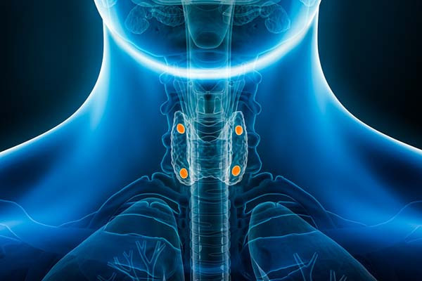
From the name, you might assume the role of the parathyroid glands is related to that of the thyroid gland. Well, you’d be wrong. The name comes solely from their location: they sit just behind the thyroid gland: two on the right side, two on the left.
The parathyroid glands are small (the size of peas), and can weigh less than a thousandth of an ounce each. Although it’s normal to have four parathyroid glands, about 13% of people have fewer and 5% have more. And some people have parathyroid glands in other locations, such as alongside the esophagus or in the chest. This variation rarely matters, unless surgery is necessary to remove one or more of them.
What do the parathyroid glands do?
Logically enough, parathyroid glands make parathyroid hormone (PTH). And what does PTH do? It has several functions, including:
- Regulating calcium: Calcium is a mineral with many essential roles throughout the body, such as maintaining bone strength, allowing nerves and muscles to function normally, and making sure blood clots as it should. Higher levels of PTH lead to higher calcium levels in the blood through actions on the kidneys and bones.
- Regulating phosphorus: Among other roles, this mineral is a key component of our DNA, bones, and teeth. Phosphorus activates essential enzymes throughout the body, including enzymes necessary for cell reproduction and survival. It also helps with nerve and muscle function.
- Regulating vitamin D: This vitamin is actually a hormone that helps maintain normal levels of calcium throughout the body, by controlling how much gets absorbed from food in the intestinal tract and how much is lost by the kidneys in your urine. Remember PTH? Well, PTH regulates production of the enzyme that converts inactive vitamin D to an active form that helps your gut absorb calcium and reduces the loss of calcium in urine.
PTH released by the parathyroid glands helps keep each of these nutrients in balance. For example, if your blood calcium level falls, your parathyroid glands make more PTH. Higher amounts of PTH prompt bones to release stored calcium into the bloodstream, and also signal the kidneys to pull back on the amount of calcium lost through urine.
What if your blood calcium level rises? Then the parathyroid glands make less PTH, which helps to correct the blood calcium level.
Which diseases involve the parathyroid glands?
The most common are:
- Hyperparathyroidism: This is a condition in which the parathyroid glands make more than the normal amount of PTH. This can be due to a benign or cancerous tumor on a single gland, or due to multiple glands becoming overactive. Or it may be due to some other trigger, such as a low level of calcium in the blood, inadequate vitamin D levels, or kidney failure. When there’s too much PTH, blood calcium levels can become dangerously high and phosphorus levels fall. Surgery may be recommended to remove the overactive gland or glands.
- Hypoparathyroidism: This rare condition is diagnosed when less than the normal amount of PTH is produced. The most common causes are prior neck surgery or radiation, autoimmune disease, or low magnesium levels.
- Parathyroid cancer: Risk factors for parathyroid cancer include certain genetic diseases and prior radiation to the neck.
Why do we rarely hear about the parathyroid glands?
The reason is that most of the time they do their job without fuss or fanfare. Although disorders of the parathyroid glands are not rare, they are just uncommon enough that most people will never hear about them. I think of the parathyroid glands as one of many parts of our bodies that play a huge role in our health, but go unappreciated because they are so good at what they do.
Many other quiet heroes, including the thymus gland, serve as testaments to the remarkable design and function of the human body. Then again, I can think of a few body parts that could be considered expendable.
The bottom line
I hope it’s comforting to know that your amazing parathyroid glands are keeping tabs on your calcium levels and helping your bones, nerves, muscles, and other organs to function normally.
Ounce for ounce, the parathyroid glands may be the most important glands you’d never heard of. Until now.
About the Author

Robert H. Shmerling, MD, Senior Faculty Editor, Harvard Health Publishing; Editorial Advisory Board Member, Harvard Health Publishing
Dr. Robert H. Shmerling is the former clinical chief of the division of rheumatology at Beth Israel Deaconess Medical Center (BIDMC), and is a current member of the corresponding faculty in medicine at Harvard Medical School. … See Full Bio View all posts by Robert H. Shmerling, MD